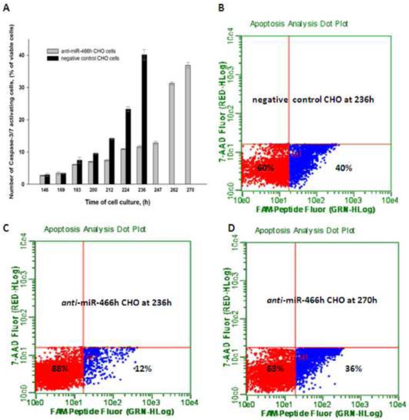
Figure 3.
Activation of Caspase-3/7 in anti-miR-466h and negative control CHO cells. (A) Time course of Caspase-3/7 activation in both cell lines (B), (C) and (D) FACS images of the ratio of viable cells negative for Caspase-3/7 activity (shown in red) and viable cells positive for Caspase-3/7 activity (shown in blue). (B) Negative control CHO at 236h. (C) Anti-miR-466h CHO at 236h. (D) Anti-miR-466h CHO at 270h.

