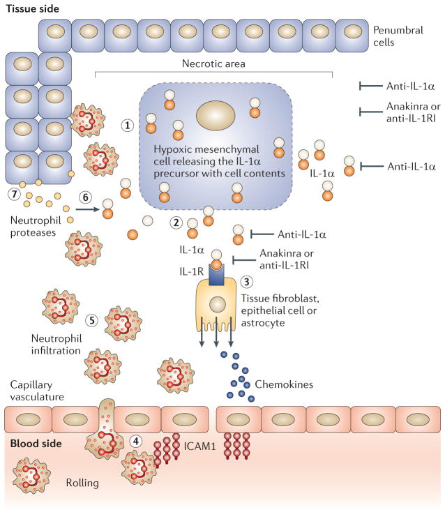
Figure 1. Initiation of sterile inflammation by IL-1α following an ischaemic event.
Step 1: in the necrotic area, dying cells lose membrane integrity. Step 2: dying cells release their contents, including the interleukin-1α (IL-1α) precursor. Anti-IL-1α antibodies neutralize IL-1α at this step. Step 3: IL-1α binds to IL-1 receptor type I (IL-1RI) on nearby resident fibroblasts, epithelial cells or in brain astrocytes, releasing chemokines and establishing a chemokine gradient. Anakinra or anti-IL-1RI antibodies block this step. The chemokine gradient facilitates the passage of blood neutrophils into the ischaemic area. Step 4: capillaries in the ischaemic tissues express intercellular adhesion molecule 1 (ICAM1). Circulating blood neutrophils roll on the endothelium, adhere to ICAM1 and enter the ischaemic tissue via diapedesis. Step 5: the number of neutrophils in the area of the necrotic event increases; the presence of local IL-1 prolongs the survival of neutrophils at this step. Step 6: neutrophil proteases cleave the extracellular IL-1α precursor into mature, more active forms. Step 7: neutrophils scavenge dying cells and release proteases that contribute to the destruction of penumbral cells.

