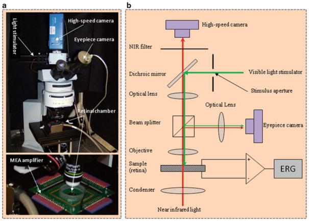
Fig. 1.
Photograph (left) and optical diagram (right) of the near infrared (NIR) light microscope for intrinsic optical signal (IOS) imaging. During measurements, isolated frog retina is illuminated continuously by the NIR light. The visible light stimulator is used to produce a visible light flash for retinal stimulation. A MEA system is used for concurrent electroretinogram (ERG) measurement of retinal activation. The dichroic mirror reflects visible stimulus light and passes the NIR recording light. The eyepiece camera is used to adjust visible light stimulus aperture at the retina. In order to ensure light efficiency for intrinsic optical signal imaging, the beam splitter is removed from the optical path after the visible light stimulator is adjusted. The NIR filter before the high-speed camera is used to block visible stimulus light, and allow the NIR probe light to reach the detector for recording stimulus-evoked IOSs. (This figure is modified from Yao (13)).

