Figure 2.


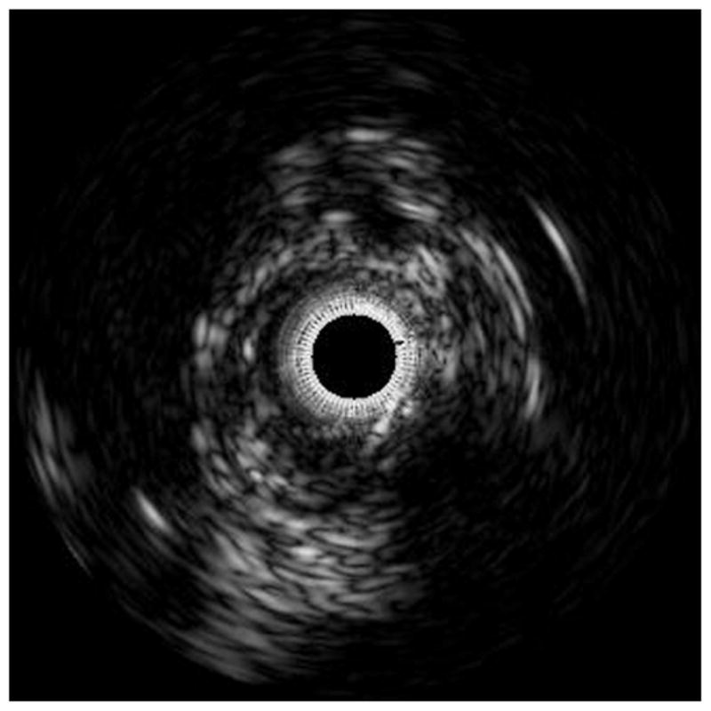
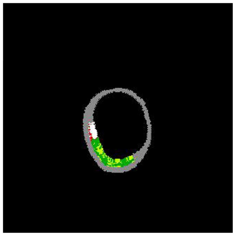
The accompanying figures demonstrate the ability to predict the composition of plaques using IVUS without any a priori information on the histology of this vessel. Figure 2a shows a gray-scale IVUS image from an anterior tibial artery. Figure 2b is the virtual histology using a modified coronary artery algorithm classified by color indicating calcium (white), necrosis (red), and fibrous (green) plaque morphology.
