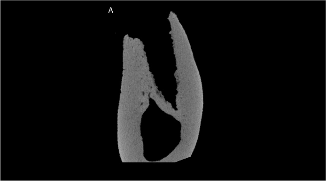
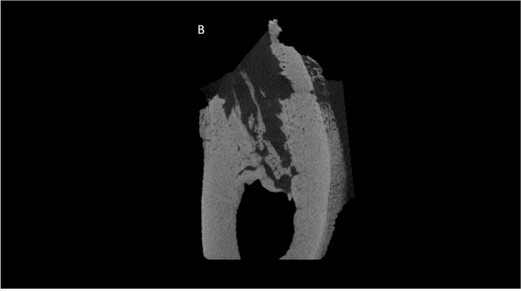
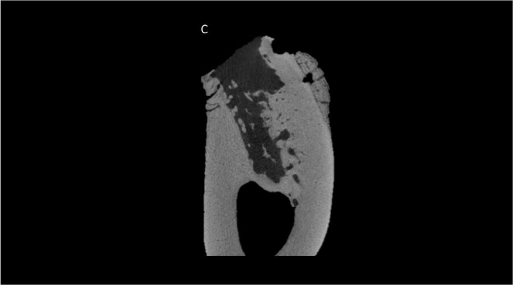
Figure 1.
Micro-computed tomography assessment of the extraction site in a zoledronate-treated animal (A) and each of the two zoledronate-treated animals that had abnormal healing (B and C). Images represent the mid-socket cross-section at 8 weeks post-extraction. Clear morphological differences can be observed between those that have abnormal healing compared to the zoledronate-treated animals including lack of alveolar socket woven bone, highly irregular/scalloped surfaces, and intense periosteal bone formation on the buccal surface.


