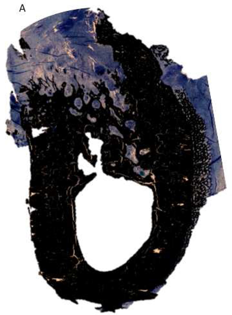
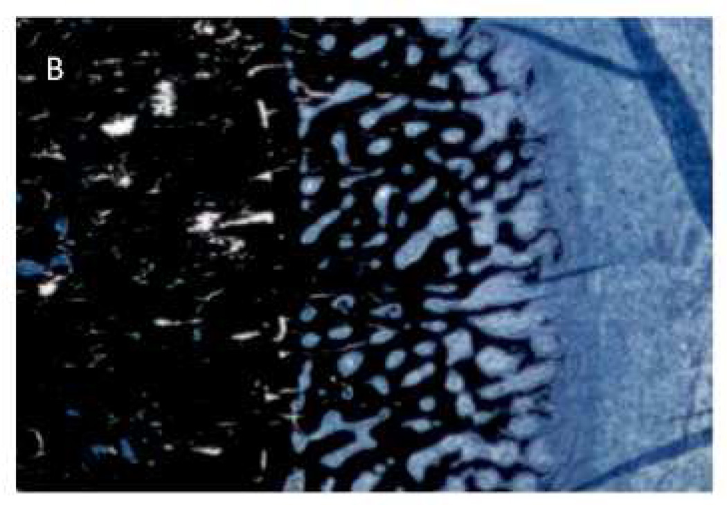
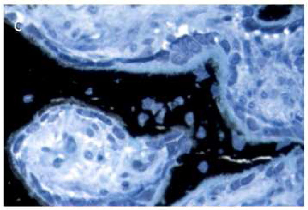
Figure 2.
Histological appearance of non-healing socket in zoledronate-treated animal. Consistent with the CT images, the histological section revealed dramatic destruction of the alveolar bone (A) and dramatic periosteal bone formation (A,B,C). The periosteal bone was continuing to form even 8 weeks post-extraction as evident by the osteoblasts and osteoid (C).


