
Figure 5.
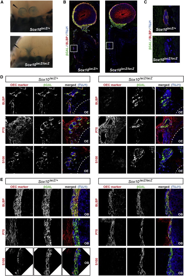
OEC Defect in the E14.5 Sox10 Mutant Mice
(A) Whole-mount X-Gal staining of the head (facial view) in heterozygous (Sox10lacZ/+, left panel) and homozygous (Sox10lacZ/lacZ, right panels) mutant embryos. The skin was removed for visualization of the olfactory bulbs, indicated by the black arrowheads.
(B) General overview of the olfactory system upon triple labeling for β-galactosidase (green), the neuronal marker (TUJ1; blue), and the OEC marker (BLBP; red), in Sox10 heterozygous (left panel) and homozygous (right panels) mutant embryos.
(C) Higher magnification of the regions boxed in (B) shows the presence of OECs ensheathing the vomeronasal nerve fibers in the heterozygous Sox10 mutant embryos and the absence of these cells in the homozygous embryos.
(D and E) Higher magnification of the nasal mesenchyme (D) or ONL (E) immunostained for an OEC marker (BLBP, P75, or S100; red), β-galactosidase (green), and the neuronal marker (TUJ1; blue), as indicated in the figure, in Sox10 heterozygous (left panels) and homozygous (right panels) mutant embryos.
Abbreviations are as follows: OB, olfactory bulb; and OE, olfactory epithelium.