
Figure 6.
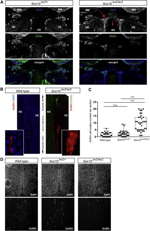
Abnormal Nerve Fasciculation, Axonal Pathfinding, and GnRH Cell Migration in Sox10 Mutant Mice
(A) TUJ1 (upper panel) and β-galactosidase (middle panel) immunostaining and a merged image (lower panel) over the nasal septum at a similar level of heterozygous (Sox10lacZ/+) and homozygous (Sox10lacZ/lacZ) mutant E14.5 embryos, as indicated. The arrow and arrowhead in the homozygote indicate the defasciculation of sensory axons and their misrouting over the nasal septum, respectively.
(B) GnRH cells along the vomeronasal nerve trajectory in wild-type (left panel) and homozygous Sox10 mutant (right panel) E14.5 embryos. The GnRH immunostaining is in red, DAPI staining is in blue, and β-galactosidase immunostaining is in green. Insets show higher magnifications.
(C) Box plots showing quantification of the number of GnRH cells along the trajectory of the vomeronasal nerve in wild-type, heterozygous, and homozygous mutant mouse embryos. Each dot corresponds to the number of GnRH-positive cell bodies counted on one side of a section. The top and bottom of each box represent the 25th and 75th percentiles, respectively. The middle line is the median. Statistical significance was tested with a Student’s t test. ***p < 0.0001. The following abbreviation is used: ns, not significant.
(D) DAPI (upper panel) and GnRH (lower panel) staining of sections at the level of the preoptic area in the wild-type (left), heterozygous (center) and homozygous (right) E14.5 embryos.
Abbreviations are as follows: OB, olfactory bulb; OE, olfactory epithelium; NS, nasal septum; and VNO, vomeronasal organ.