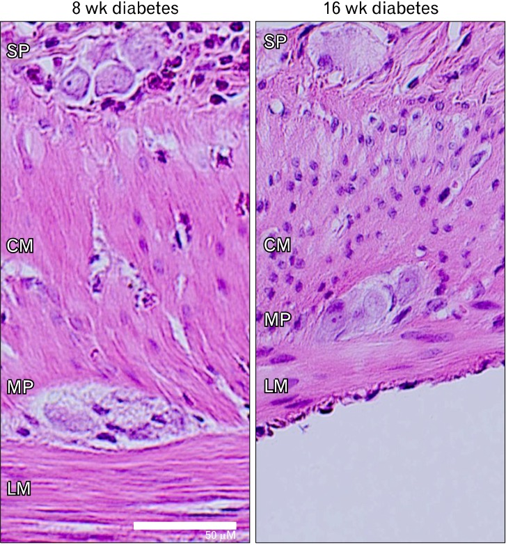
Figure 3.
Representative image of 8 and 16 weeks diabetic rat by H&E staining. Eight weeks after onset of diabetes, infiltration of polymorphonuclear leukocyte cells into enteric nervous system (SP, submucosal plexus; MP, myenteric plexus) was present with thickening of the muscular layers (CM, circular muscle; LM, longitudinal muscle). However, 16 weeks after onset of diabetes, no inflammatory infiltrate was seen. Scale bar = 50 µm.

