
Figure 3.
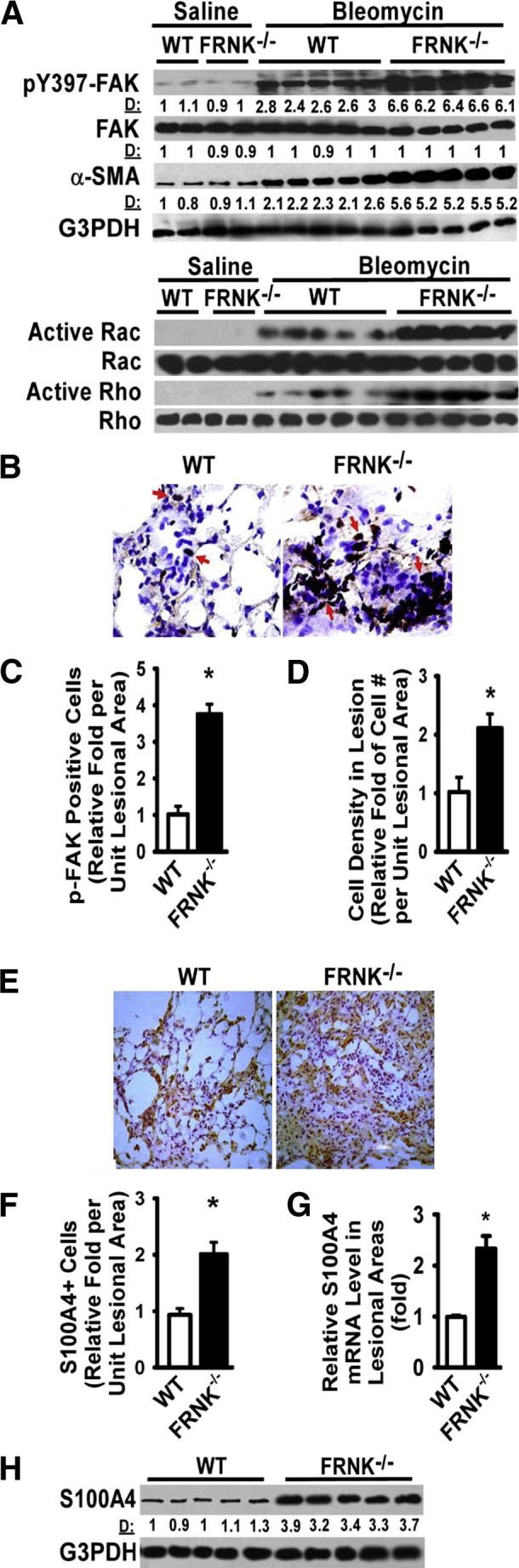
FRNK deficiency potentiates integrin/migration signaling and myofibroblast differentiation in fibrotic lungs. A: FRNK−/− and WT lungs were harvested at day 14 after bleomycin or saline instillation and lysed, and equivalent amounts of whole lung lysate were examined for indicated signaling and α-SMA expression. D: Relative band density by densitometry. B: Lesional cells containing active FAK (pY397-FAK) were detected by IHC on frozen lung tissue sections from WT and FRNK−/− mice. Arrows indicate pY397-FAK–positive cells. Original magnification, ×400. C: Lesional pY397-FAK–positive cells were quantified as the fold of pY397-FAK–positive cells in FRNK−/− mice relative to that in WT mice. D: The overall cell density in lesions was determined. E: S100A4-positive cells in fibrotic lesions were detected by IHC (brown). F: Lesional S100A4-positive cells were quantified as the fold of S100A4-positive cells in FRNK−/− mice relative to that in WT mice. G: Fibrotic/lesional tissues were captured by LCM from frozen lung sections. Total RNA was extracted, and the level of S100A4 mRNA was determined by quantitative real-time PCR (normalized to GAPDH) and represented as relative fold to that in WT mice. H: The S100A4 protein level in fibrotic lungs of WT and FRNK−/− mice was determined by using Western blot analysis. Data were represented as means ± SE (n = 8 animals per group). ∗P < 0.01.