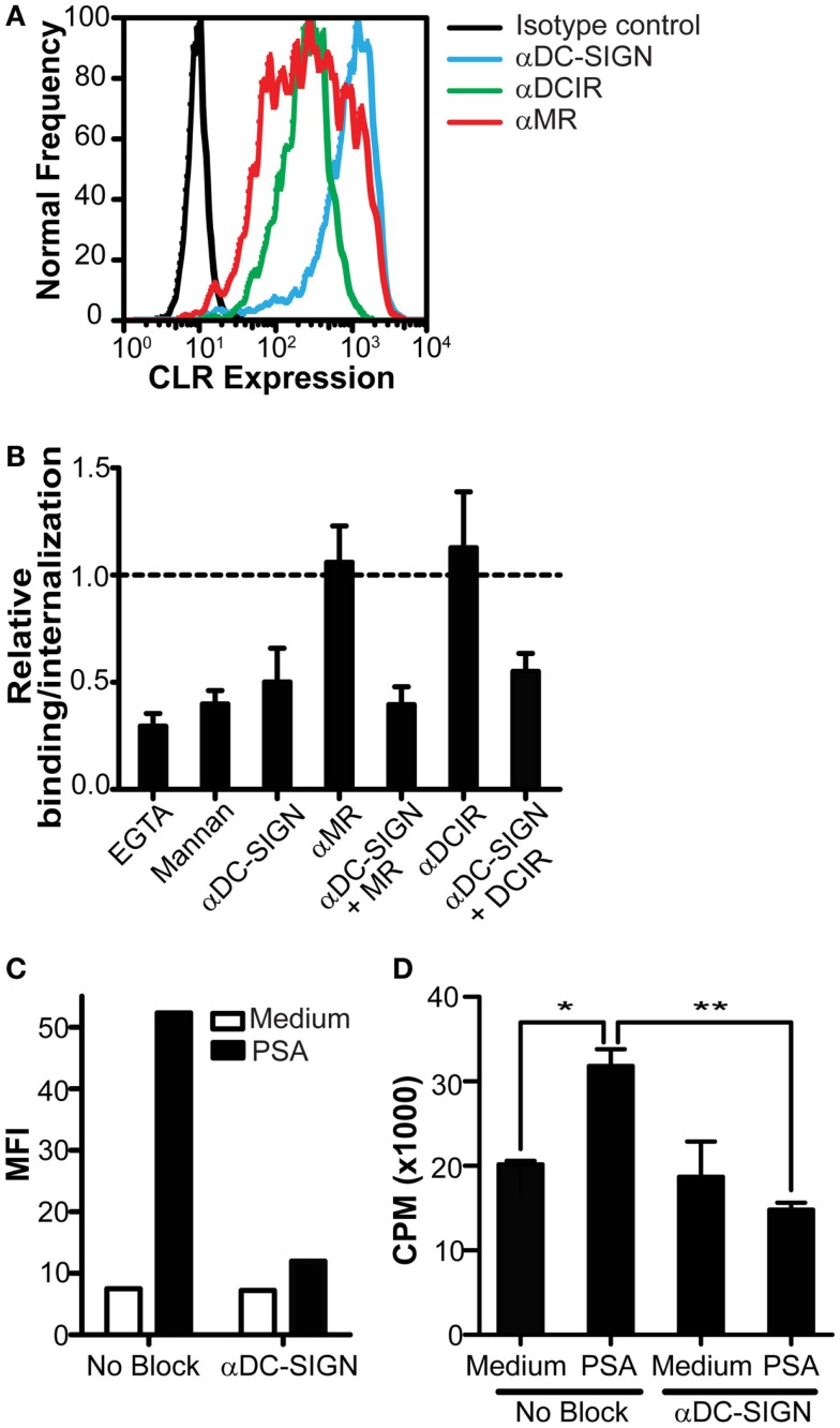
Figure 3.
Inhibition of DC-SIGN function decreases PSA internalization and PSA-induced T cell proliferation. (A) Expression of DC-SIGN, MR, and DCIR on DCs as measured by flow cytometry. Black line represents isotype control, blue line indicates DC-SIGN expression, green line indicates DCIR expression, and red line represents MR expression. (B) PSA-binding/internalization by DCs is only blocked in the presence of a DC-SIGN blocking antibody. Data is shown relative to DCs incubated with PSA in the absence of inhibitors. (C) DC-SIGN blocking antibodies decrease PSA-binding/internalization by DCs. Binding/internalization of PSA-AF488 by irradiated DCs was measured by flow cytometry after 3 h incubation at 37°C. (D) Inhibition of DC-SIGN function decreases PSA-specific T cell proliferation. Irradiated DCs were pre-incubated with a DC-SIGN blocking antibody and subsequently incubated with PSA for 3 h at 37°C, extensively washed, and incubated with autologous CD4+ T cells in the presence of the NO donor glyco-SNAP-2. T cell proliferation was measured by [3H]-thymidine incorporation. Data is shown as mean ± SD of triplicates (*p < 0.05 **p < 0.01). All experiments were performed three times with independent donors, one representative experiment is shown.

