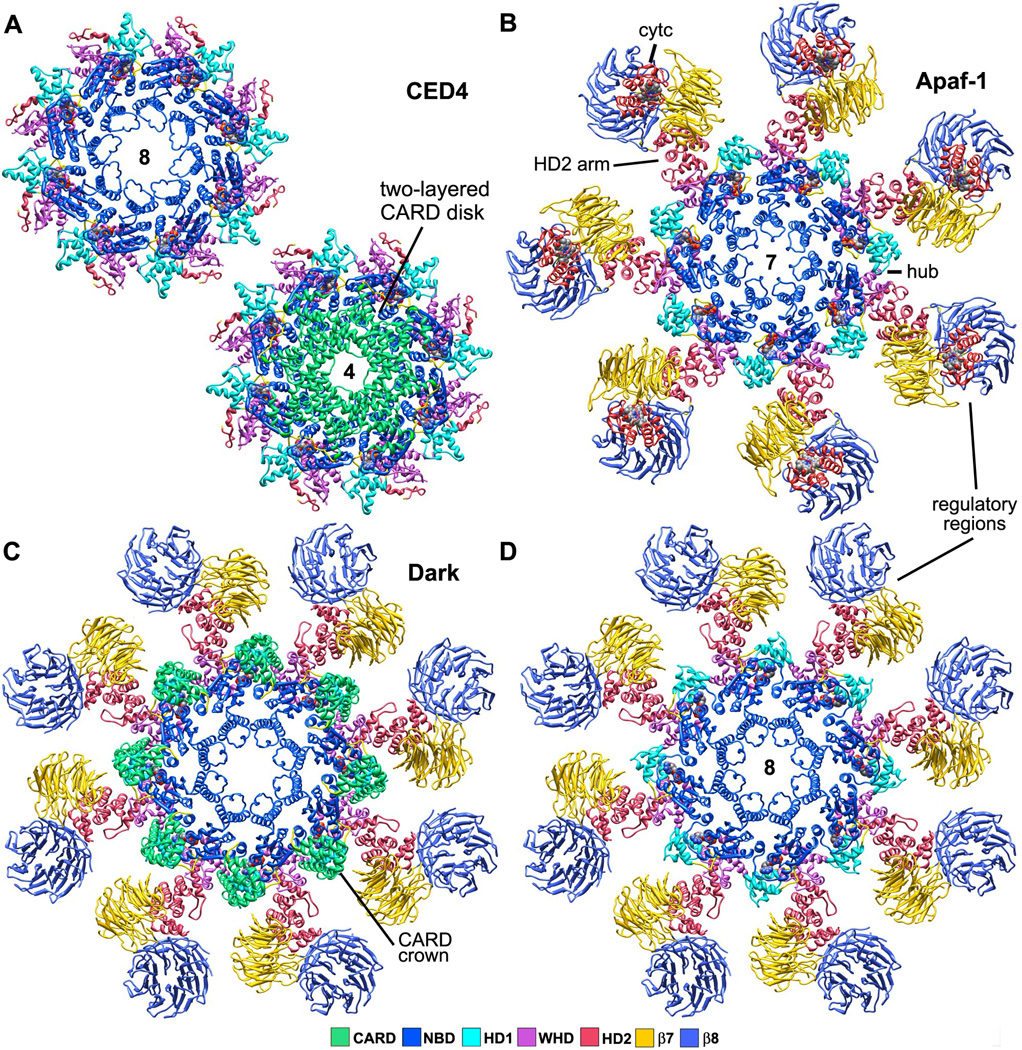
Figure 3. Top views of worm, human and fly apoptosomes.
A. (top left) The central hub of the C. elegans apoptosome has quasi 8-fold symmetry. (lower right) CARDs from the A and B monomers of CED-4 form two tetrameric rings stacked along the 8-fold axis of the hub to create an apoptosome with overall 4-fold symmetry (Qi et al., 2010; 3LQQ).
B. The human apoptosome contains seven Apaf-1 molecules whose CARDs are disordered in the ground state (Yuan et al., 2010; submitted).
C. A Drosophila single ring apoptosome is shown with eight CARDs that bind to the lateral surface of their respective NBDs to form a crown (Yuan et al., 2011a; 1VT4, 3IZ8).
D. The CARD crown has been removed to show the similarity of the octagonal fly apoptosome and the heptameric Apaf-1 apoptosome in top views (compare with panel B). Note that mammalian cytochrome c does not bind to Dark in these complexes.

