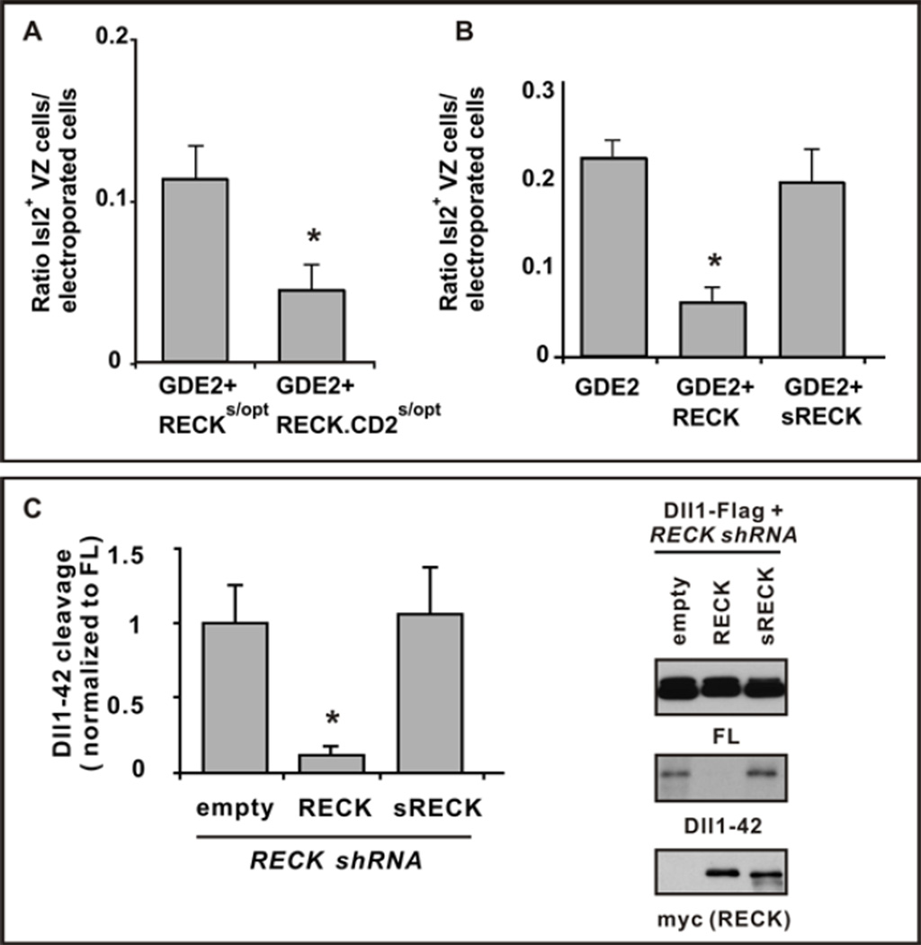
Figure 4. GDE2 inactivates RECK by GPI-anchor cleavage.
(A, B) Graphs quantifying ratio of ectopic Isl2+ VZ MNs normalized to the number of transfected GDE2 cells. Mean ± s.e.m., two-tailed t-test, (A) Suboptimal levels of plasmids expressing RECKs/opt or RECK-CD2 s/opt were coelectroporated with GDE2. RECK-CD2 was more effective than RECK in suppressing GDE2 dependent MN generation, *p= 0.0306 (n=5); (B) Plasmids expressing RECK or sRECK were coelectroporated with GDE2; RECK effectively suppressed GDE2 function but sRECK did not, *p= 5.59×10⁻⁵ (n=8–10) compared with GDE2. (C) Western blot of extracts of chick spinal cords electroporated with Dll1-Flag and RECK shRNA targeting 3’ UTR to detect full-length (FL) and processed Dll1-42. The phenotype is rescued by exogenous plasmids expressing WT RECK ORF but not sRECK. Densitometric quantification of Dll1-42, mean ± s.e.m., n=4. Two-tailed t-test, *p=0.013 compared with empty.

