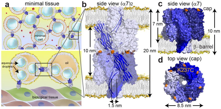
Figure 1. An engineered αHL pore dimer.

(a) Minimal tissue models. Upper panel: minimal cells communicating through protein conduits that span both bilayers (upper panel). Lower panel: monolayer-encased droplet networks8 interconnected and linked to biological tissues through protein conduits. (b) Space-filling model of the engineered pore dimer, (α7)2, in which two α7 units (the normal form of the αHL pore) are linked cap-to-cap, and can span two bilayers simultaneously. (c) The α7 units were covalently attached through disulfide bonds between cysteine residues (orange) at position 237 in the αHL K237C mutant. The cysteine residues are located on the cap of the α7 heptamer, one on each subunit. (d) Top view of the cap and the location of the cysteine residue at position 237 (orange) in α7. The space filling models of α7 (PDB 7AHL) and (α7)2 were created in PyMOL.
