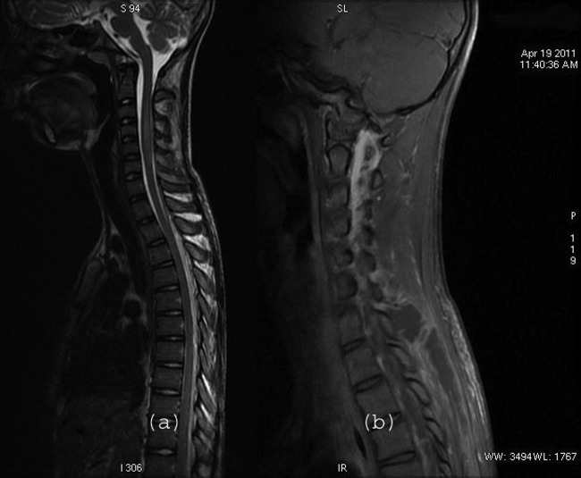
Figure 1.
T2-weighted sagittal image showing epidural collection dorsally extending from D1 to the dorsolumbar region with cord oedema in the cervical region from the C2–3 to the C6 level. Contrast-enhanced fat suppressed T1-weighted sequence in the right parasagittal plane showing an abscess appearing as a necrotic collection in the posterior paraspinal muscles with enhancing capsule/granulation tissue peripherally and involvement of the C6–7 apophyseal joint.

