
Figure 2.
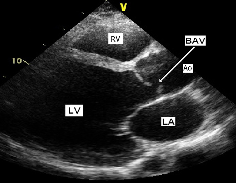
Transthoracic echocardiogram of the proband (subject ‘B’) showing a severely dilated left ventricle (dimensions of 9.9 cm in diastole and 8.9 cm in systole, normal range 4.2–5.9 cm) and BAV (white arrow) with a typical off-midline closure of leaflets. Left ventricular ejection fraction 14% (normal >55%). Ao, aorta; BAV, bicuspid aortic valve; LA, left atrium; LV, left ventricle; RV, right ventricle.