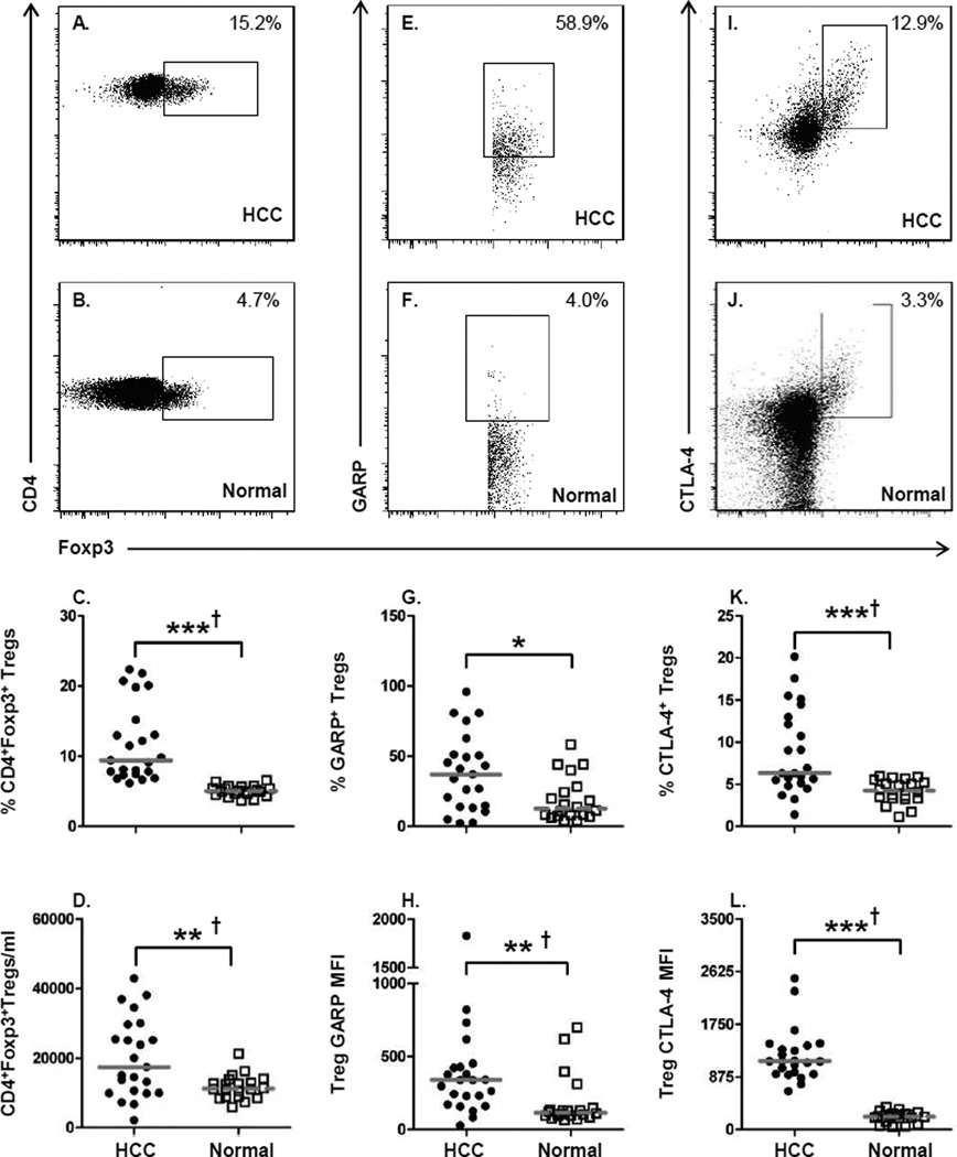
Figure 1. Increased numbers of GARP and CTLA-4 expressing Tregs in HCC patients.
Flow cytometric analysis was performed on PBMCs from HCC patients (n=23) and healthy
controls (n=20). (A, B) Representative staining from an individual HCC patient and
normal healthy donor for the frequency of CD3+CD4+Foxp3+ T cells.
(C) Frequency and (D) absolute number of cells/ml of
CD4+Foxp3+ Tregs in peripheral blood of HCC patients and normal healthy
subjects. (E, F) Representative staining of GARP on
CD3+CD4+Foxp3+ T cells. (G) Frequency of
GARP+ Tregs and (H) GARP expression levels measured by mean fluorescent
intensity (MFI) on Tregs. (I, J) Representative staining of CTLA-4 on
CD3+CD4+Foxp3+ T cells. (K) Frequency of
CTLA-4+ Tregs and (L) CTLA-4 expression levels. Each symbol represents an
individual HCC patient ( ) or normal healthy subjects (
 ); lines represent median
values for the group. * P < 0.05, ** P < 0.01, ***
P < 0.001, Mann-Whitney U test; † P < 0.05
Hochberg adjustment for multiple comparison.

