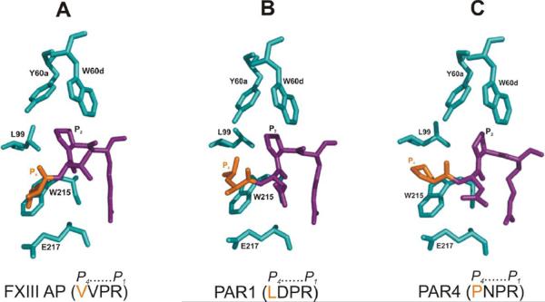
Figure 2. Conformational features associated with segments of FXIII AP, PAR1, and PAR4 bound to the active site of thrombin.
(A) Selected residues of human thrombin that surround FXIII AP (34–37), (B) selected residues of human thrombin that surround PAR1 (38–41), and (C) selected residues of murine thrombin that surround murine PAR4 (56–59). The X-ray PDB codes include 1DE7 (human), 3LU9 (human), and 2PV9 (murine), respectively. For each panel, the thrombin residues are in cyan, P4 residue of the substrate is in orange, and the P3-P1 residues of the substrate are in purple.

