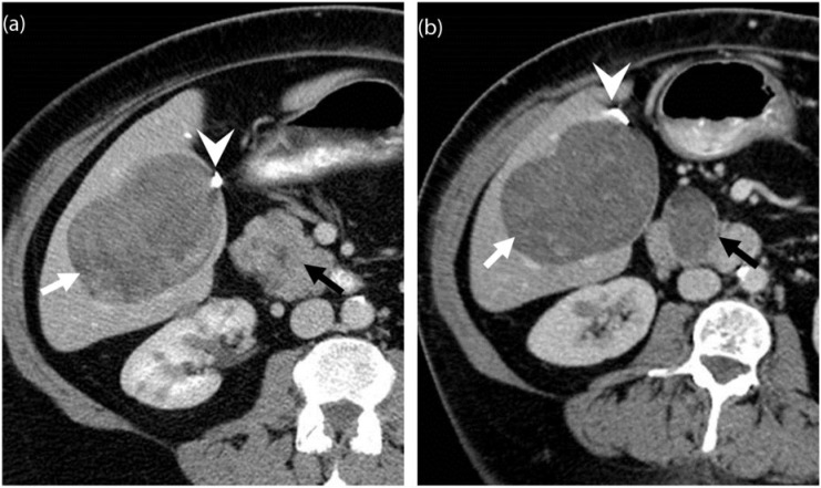
Figure 9.
A 51-year-old woman with metastatic myxoid liposarcoma. (a) Axial contrast-enhanced CT image of the abdomen demonstrates a large heterogeneous metastatic mass in the liver (white arrow) and the pancreas (black arrow). Note the surgical clip related to previous cholecystectomy (arrowhead). Note the incidental renal cortical cysts. (b) Axial contrast-enhanced CT image 6 months after treatment with trabectedin reveals a mild increase in the size of both the lesions with homogeneous decreased internal density suggestive of treatment effect (white and black arrows). The increase in size represents pseudo-progression rather than true progression, wherein a lesion may increase in size but is in fact responding to therapy as shown by the density changes.

