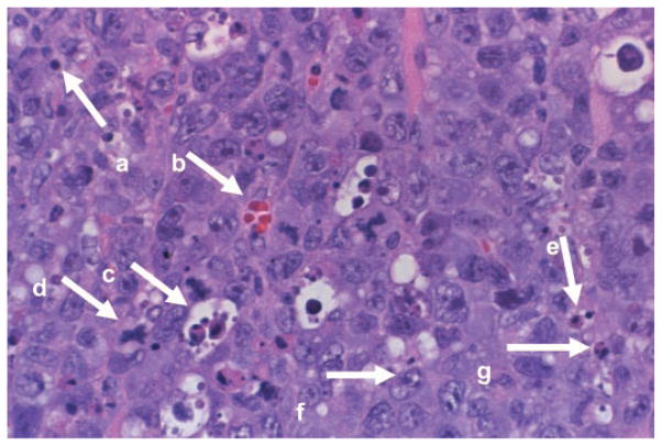
Fig. 1.
A representative hematoxylin and eosin-stained slide (×400 magnification) from a subcutaneous EL-4 tumor from a control mouse. Individual components of the tumor were identified by a blinded observer. a, Lymphocyte; b, blood vessel; c, macrophage (with ingested cellular debris); d, mitotic figure; e, apoptotic body; f, EL-4 cell; g, neutrophil.

