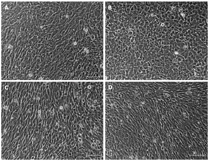
Figure 2.

Phase-contrast microscopic images of primary intestinal myofibroblasts, mouse embryonic fibroblasts, LmcMF, and SmcMF. A: Primary intestinal myofibroblasts; B: Mouse embryonic fibroblasts; C: LmcMF; D: SmcMF were cultured to confluence, and the phase-contrast microscopic images were taken. Representative images are shown. Scale bars indicate 200 μm.
