
Figure 6.
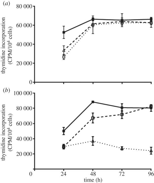
Proliferation of (a) HUES7 and (b) HUES1 cells on fibronectin (filled squares), the 120 (open squares) and the 70 kDa (triangles) fragments measured using the uptake of [3H] thymidine. The cells were incubated over a time course of 24, 48, 72 and 96 h and pulsed with 1 mCi [3H] thymidine for the final 4 h of culture at each time point. Triplicate experiments were run.