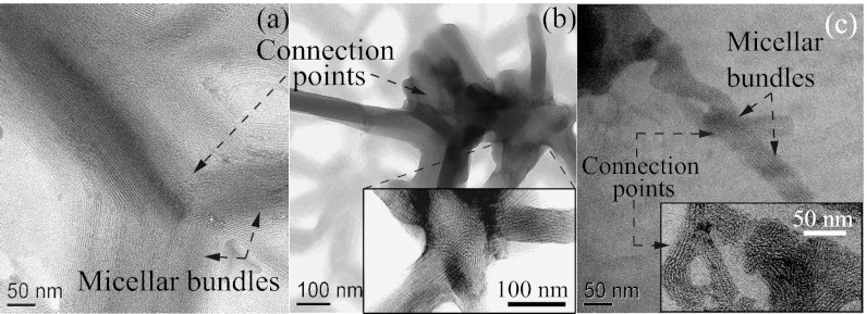
Fig. 5.
(A and B) TEM images of FISP50 show branched, entangled, and multiconnected micellar bundles. (C) TEM image of FISP100 shows entangled, branched, and multiconnected micellar bundles. (Insets) Zoomed-in images where branching points and connection points are presented. TEM was conducted with a Tecnai T-12 transmission electron microscope at 120 kV.

