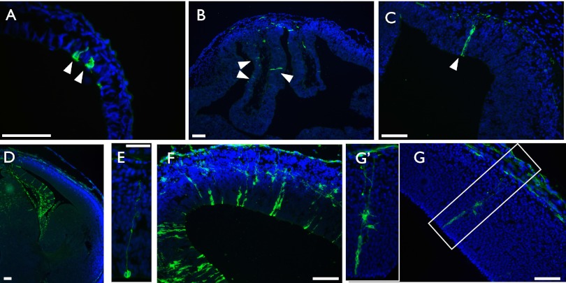
Fig. 1.
Axin2CreERT2 marks cells of the neural lineage at multiple developmental time points. Pregnant females carrying Axin2CreERT2/+; R26RmTmG/+ embryos were injected with tamoxifen at various developmental time points and embryos examined 24–48 h later. (A) E6.5–E7.5; (B and C) E8.5–E10.5; (D and E) E12.5–E13.5; and (F and G) E12.5–E14.5. (A) DAPI-stained tissue section of an Axin2CreERT2/+; R26RmTmG/+ embryo at E7.5, 24 h after administration of tamoxifen shows rare GFP+ cells in the ectoderm. (B) An E8.5–E10.5 trace shows GFP+ cells in the pallium and midline structures (C), which are beginning to take on elongated radial glial cell morphology. (D) In an E12.5–E13.5 traced forebrain, many GFP+ cells are present in the cortical hem and septum, and fewer in the pallium, and none in the ganglionic eminences. (E) Pallial GFP+ cell with a long radial fiber undergoing cell division at the ventricular surface, traced E12.5–E13.5. (F) After an E12.5–E14.5 trace, there is an increase in the total number of GFP+ cells in a given clone compared with E12.5–E13.5, although overall regional distributions remain the same. (G) E12.5–E14.5, a single clone of GFP+ cells containing at least three radial glial cells and multiple differentiated progeny. Boxed region is magnified in the Inset (G′). (A) Transverse section, (B–G) coronal section. DAPI staining is shown in blue, GFP staining is shown in green. (Scale bar, 50 μm, except in E and Inset in G, where it represents 25 μm.)

