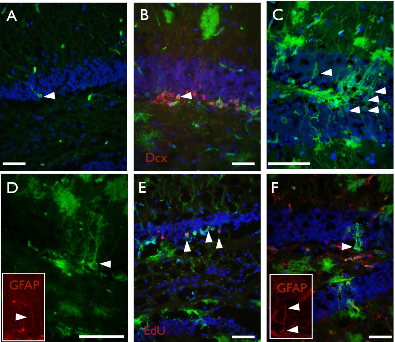
Fig. 5.
Axin2CreERT2 labels stem cells of the dentate gyrus. Representative images of brain sections from Axin2CreERT2/+; R26RmTmG/+ traced postnatally for various lengths of time. (A) P14–16, 2 d after tamoxifen, rare GFP+ cells are found in the subgranular zone, but no neurons are labeled. (B) P14–P81, GFP+ Dcx+ cells are present, as well as differentiated neurons (D) and subgranular cells that are GFP+ GFAP+ (E) (Inset shows GFAP staining). (F) P14–P65, GFP+ EdU+ cells can be found in the subgranular zone 30 d after EdU administration. (G) P14–P379, 1 y after tamoxifen, GFP+ GFAP+ cells are still present (Inset shows GFAP staining). DAPI is shown in blue, GFP is shown in green, and all other markers are shown in red. Arrowheads point to cells of interest and double positive cells. (Scale bar, 50 μm.)

