Fig. 1.
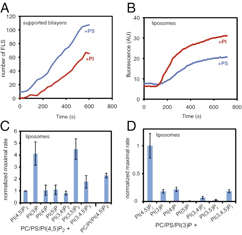
PI and PI(3)P contribute to actin polymerization from liposomes. (A) Numbers of filopodia-like structures from supported lipid bilayers showing that filopodia-like structure formation is favored by PS and not PI. The lipid compositions were 48% PC, 48% PS or PI, and 4% PI(4,5)P2. (B) Pyrene actin assay of actin polymerization from liposomes showing that it is favored by PI rather than PS, with the same liposomes used in A. (C) Bar chart showing the maximal rate of actin polymerization (slope of a pyrene actin assay) with a lipid composition of 48% PC, 47% PS, 4% PI(4,5)P2, and 1% of the test phosphoinositide, or PC/PI/PI(4,5)P2. The values are normalized against PC/PS/PI(4,5)P2 liposomes. (D) Substituting PI(4,5)P2 and keeping 1% PI(3)P constant shows that PI(4,5)P2 is essential. Data are normalized to PC/PS/PI(3)P/PI(4,5)P2 liposomes. Data in A and B is representative of four independent experiments. Data in C and D is the average of three experiments, with error bars showing the SD.

