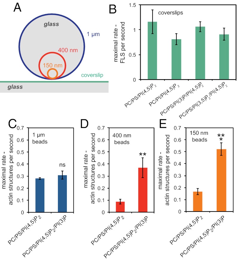
Fig. 3.
Membrane curvature contributes to actin polymerization from PI(3)P/PI(4,5)P2 membranes. (A) Relative sizes of glass beads (to scale) compared with a coverslip. (B) Bar chart showing that the rate of appearance of filopodia-like structures is unaltered by the presence of PI(3)P or PI(3,5)P2. The lipid compositions were 48% PC, 47% PS, 1% PI(3)P or PI(3,5)P2, and 4% PI(4,5)P2. (C) Rate of appearance of actin structures growing on bilayers composed of 48% PC, 48% PS, 4% PI(4,5)P2 and 48% PC, 47% PS, 4% PI(4,5)P2, and 1% PI(3)P supported on 1-μm-diameter glass beads showing no significance difference between the compositions. (D) Rate of appearance of actin structures growing on bilayers supported on 400-nm-diameter glass beads showing a very significant increase in the presence of PI(3)P (**P = 0.008). (E) Number of actin structures growing on bilayers supported on 150-nm-diameter glass beads showing a significant increase in the presence of PI(3)P (***P = 0.0001). Data are the mean of six experiments; error bars show SEM.

