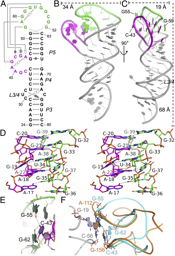
Fig. 3.
Stem I57 crystal structure. (A) Secondary structure of the stem I57 crystallization construct with tertiary contacts drawn by using the Leontis–Westhof notation (43), with the AG bulge (magenta) and distal loop (green) highlighted. (B and C) Orthogonal views of the stem I57 crystal structure shown as a cartoon, colored as in A. (D) Stereo image of the AG bulge–distal loop interaction. (E) The apical base triple, highlighted in sticks, colored as in B and C. Oxygen, nitrogen, and phosphorus are shown in red, blue, and orange, respectively. Hydrogen bonds are shown as dashed lines. Stem I57 is in the same orientation as in C. (F) Superposition of the stem I apical loops (cyan) and the RNase P specificity domain (orange), which is in complex with tRNA (gray) [Protein Data Bank (PDB) ID 3Q1Q]. Structures are shown as ribbons with interacting bases highlighted in sticks. Hydrogen bonds in the stem I apical base triple are highlighted as dashed lines.

