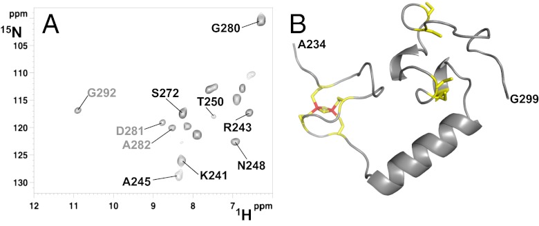
Fig. 2.
Structural characterization of [2Fe-2S]-CIAPIN1-single. (A) 1H-15N IR-HSQC-AP spectrum, optimized to detect fast relaxing 1H resonances, showing 13 backbone NH resonances of the [2Fe-2S]-CIAPIN1-single, 10 of which (in black) were completely lost in the standard diamagnetic 1H-15N HSQC experiment (the three residues also detected in the diamagnetic 1H-15N HSQC are shown in gray. (B) Representative model of the CIAPIN1 domain of anamorsin derived from a molecular dynamics simulation of 100 ns. The [2Fe-2S] cluster bound to the CX8CX2CXC motif is shown; the four cysteines coordinating the [2Fe-2S] cluster and the four cysteines of the CX2CX7CX2C motif are shown.

