Fig. 5.
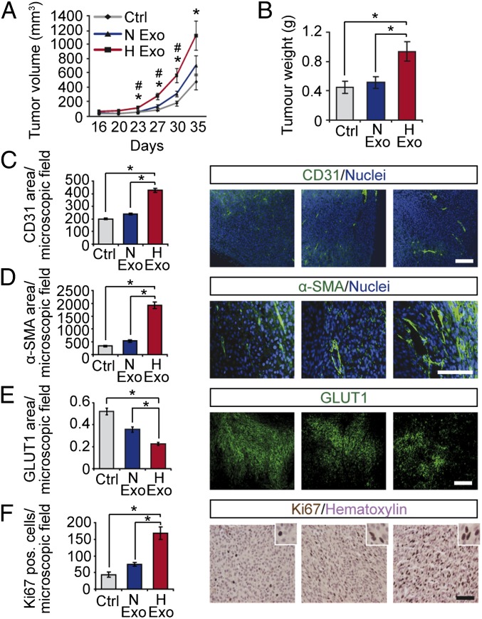
Hypoxic induction of exosome-mediated stimulation of tumor development. Human GBM xenografts were established with or without exosomes (1 μg/mL) from normoxic (N Exo) or hypoxic (H Exo) GBM cells. Tumor volumes at the indicated time points (A) and final tumor weights (B) were determined. Data are presented as the mean ± SEM, *P < 0.05 compared with untreated Control; #P < 0.05 compared with N Exo tumors (n = 5). Tumors were analyzed by immunofluorescence microscopy for vascular density (C), pericyte coverage (D), hypoxic area (E), and proliferation (F). (Scale bars: 100 µm.) Results are the mean ± SEM, *P < 0.05.

