Fig. 2.
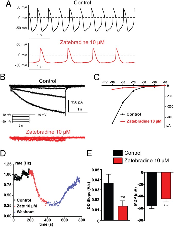
A subset of hESC-CMs exhibits prominent If-dependent pacemaker. (A–D) Combined current- and voltage-clamp recordings were performed in the same cell. (A, Upper) Spontaneous AP pattern of a hESC-CM recorded under current-clamp in control conditions. (B) The If current was subsequently recorded in the same cell, under voltage-clamp, in the absence (black traces) and presence of 10 µM zatebradine (red traces) by stepping the membrane from a holding potential of −40 mV to −90 mV in 10-mV decrements for 3 s pulse duration. (C) Current-voltage relationship of the If current from this cell showing the strong inhibitory effect of zatebradine. Once If current inhibition by zatebradine was monitored under voltage-clamp, the AP pattern of the same cell was examined under current-clamp, during continuous zatebradine exposure (see A, Lower). Note the bradycardia and the MDP depolarization. (D) Bradycardia was measured over time by the instantaneous frequency between two contiguous APs (in Hz) before (black dots), following 10 µM zatebradine (red dots), and during washout (blue dots). (E) Zatebradine (10 µM) significantly decreased the DD slope (**P = 0.0078; n = 11) and depolarized the MDP in this subset of cells (**P = 0.0058; n = 11).

