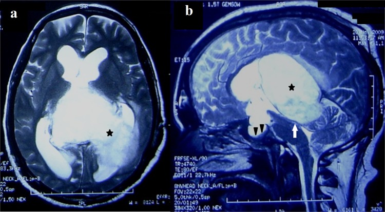
Figure 2.
T2-weighted axial and sagittal MR image shows a large hyperintense lesion in the atrium and occipital horn of the left lateral ventricle (star). Compression of the aqueduct and posterior third ventricle (arrow) is evident with dilation of the proximal part of the third and bilateral lateral ventricles. Enlarged sella and compressed pituitary gland is also seen (arrow heads).

