Figure 1.

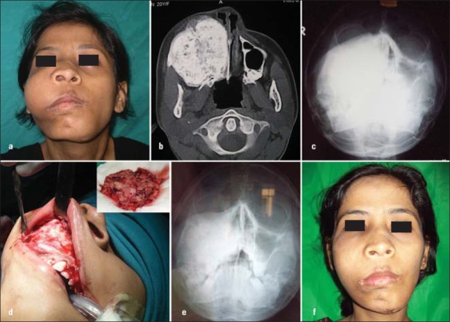
(a) Monostotic maxillary fibrous dysplasia, (b) CT showing extensive maxillary involvement, (c) PNS Radiograph showing maxillary sinus obliteration, (d) Intraoral view of the lesion - Amount of bone removed, (e) Postoperative PNS view, (f) Postoperative view
