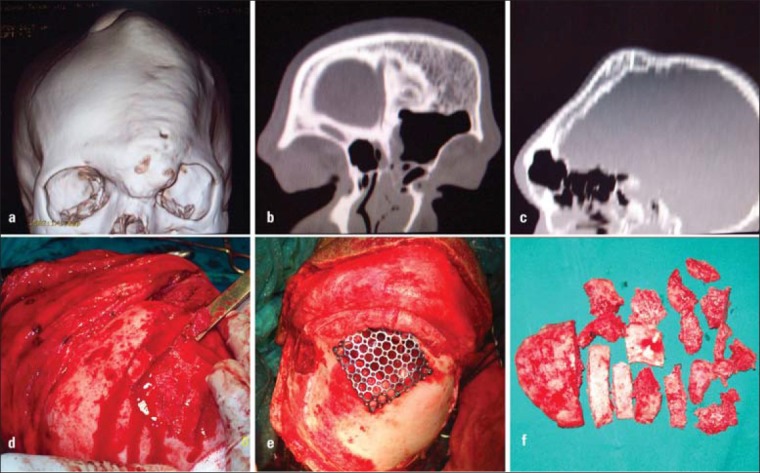
Figure 3.

(a) CT showing frontal and parietal involvement, (b) Coronal CT view, (c) Sagittal CT view, (d) Removal of bone using osteotome, (e) Reconstruction of the frontal sinus wall using mesh, (f) The bone chips that were removed during frontal contouring
