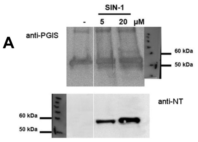
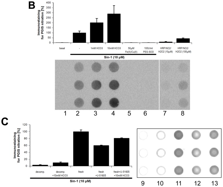
Figure 4.
Nitration of purified, recombinant PGIS by in situ generated peroxynitrite. (A) Detection of 3-nitrotyrosine in PGIS by Western blot analysis using a polyclonal amti-PGIS antibody and a monoclonal anti-3-nitrotyrosine antibody. PGIS (300 nM) was treated with peroxynitrite generated in situ from Sin-1 (5 and 20 μM). Data are representative of two independent experiments; (B) Detection of 3-nitrotyrosine in PGIS by dot blot analysis using a monoclonal anti-3-nitrotyrosine antibody. PGIS (80 nM) was not treated (lane 1) or treated with peroxynitrite generated in situ from Sin-1 (10 μM) (lane 2) in the absence or presence of 1 mM (lane 3) or 10 mM (lane 4) bicarbonate or 50 μM iron(II) plus copper (II) ions (lane 5) or 100 U/mL PEG-SOD (lane 6). PGIS was also incubated with 0.1 μM horseradish peroxidase (HRP) plus 10 μM nitrite/hydrogen peroxide (lane 7) or plus 100 μM nitrite/hydrogen peroxide (lane 8); (C) Detection of 3-nitrotyrosine in PGIS by dot blot analysis using a monoclonal anti-3-nitrotyrosine antibody. PGIS (80 nM) was treated with 10 μM decomposed Sin-1, from a 1 mM Sin-1 solution in 1 M potassium phosphate buffer pH 7.4 incubated for 90 min at 37 °C, in the absence (lane 9) or presence of 10 mM bicarbonate (lane 10), freshly prepared 10 μM Sin-1 in the absence (lane 11) or presence of 2 μM U-51605 (lane 12) as well as 2 μM U-51605 plus 10 mM bicarbonate (lane 13). All incubations were performed in 0.1 M potassium phosphate buffer pH 7.4 at 37 °C for 90 min. Data are means ± SEM of three independent experiments.


