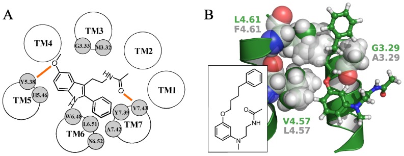
Figure 6.
(A) Representation of the binding scheme of 2-phenylmelatonin within the MT1 binding site proposed by Rivara et al. Hydrogen bond interactions are depicted with orange lines; (B) The MT1-selective phenylbutyloxy derivative (ball-and-sticks representation, with green carbons) docked within the MT1 receptor model. Residues 3.29, 4.57 and 4.61 are represented as green (MT1) and transparent light gray (MT2) spheres. The structure of the phenylbutyloxy derivative is reported in the inset.

