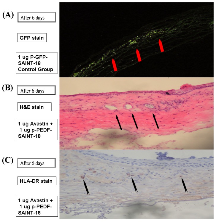
Figure 5.
The tissue was histologically evaluated on day 6 after administration. (A) GFP was expressed within the corneal intrastromal layer and keratocytes after the implantation of p-GFP-SAINT-18. No vascular lumen was noted; (B) The intervening stroma displayed cells, edema, a mononuclear inflammatory response, and numerous vascular lumens (indicated by arrows) after hematoxylin and eosin staining in the corneal and subconjunctival substantia propria (100× magnification); (C) Inflammation was detected by the presence of macrophages and other inflammatory cells, such as lymphocytes, by immunohistochemical staining (anti-rat HLA-DR antibody) in the corneal and subconjunctival substantia propria (100× magnification).

