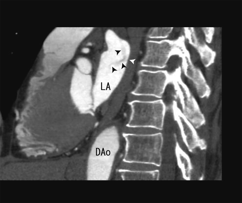
Figure 2.

The 64- multidetector CT sagittal images showing the thrombi from the left lower pulmonary vein to left atrium (white and black arrow head). The merge of the thrombi was vague. A part of the thrombi seemed to be attached to the posterior wall of LA. Dao, descending aorta, LA, left atrium.
