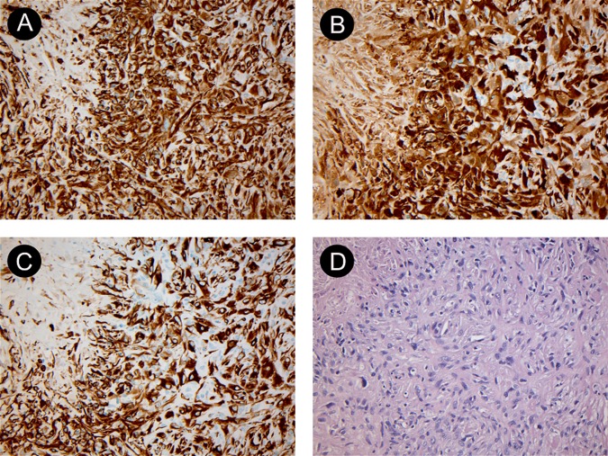
Figure 3.
Histology specimens from video-assisted thoracoscopic surgery showing biphasic malignant pleural mesothelioma. (A) shows a vimentin stain ×200, (B) shows calretinin stain ×200, (C) shows cytokeratin stain ×200 and (D) shows H&E stain ×200. The specimens were examined by the Department of Pathology at our institution and reviewed by the national reference centre for malignant pleural mesothelioma. Courtesy of Department of Pathology, Odense University Hospital.

