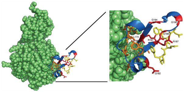
FIG. 5. Conservation of residues within the β-subdomain of CheR.

The CheR-pentapeptide structure (left) is displayed as a space-filling model (green), except for the β-subdomain (blue) and pentapeptide (gold), which are shown as a ribbon diagram and in stick representation, respectively. An enlarged view of the β-subdomain (right) shows conserved residues in stick representation. Universally conserved β-subdomain residues are shown in orange and residues that are conserved exclusively in pentapeptide-dependent β-subdomains are shown in red. Images were generated using Pymol (Delano, 2002).
