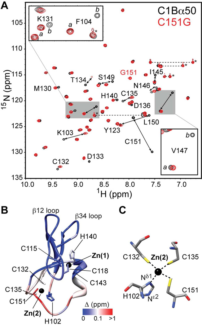
Figure 4.
C1Bα50 exists in two slowly exchanging conformations. (A) Overlay of the 15N-1H HSQC spectra of C1Bα50 and C151G variant. The insets show the expansions for Lys131, Phe104, and Val147. (B) Chemical shift perturbation Δ mapped onto the structure of C1Bα.62 The coordinates are the courtesy of Dr. U. Hommel. (C) Coordination geometry of Zn(2) showing four protein ligands.

