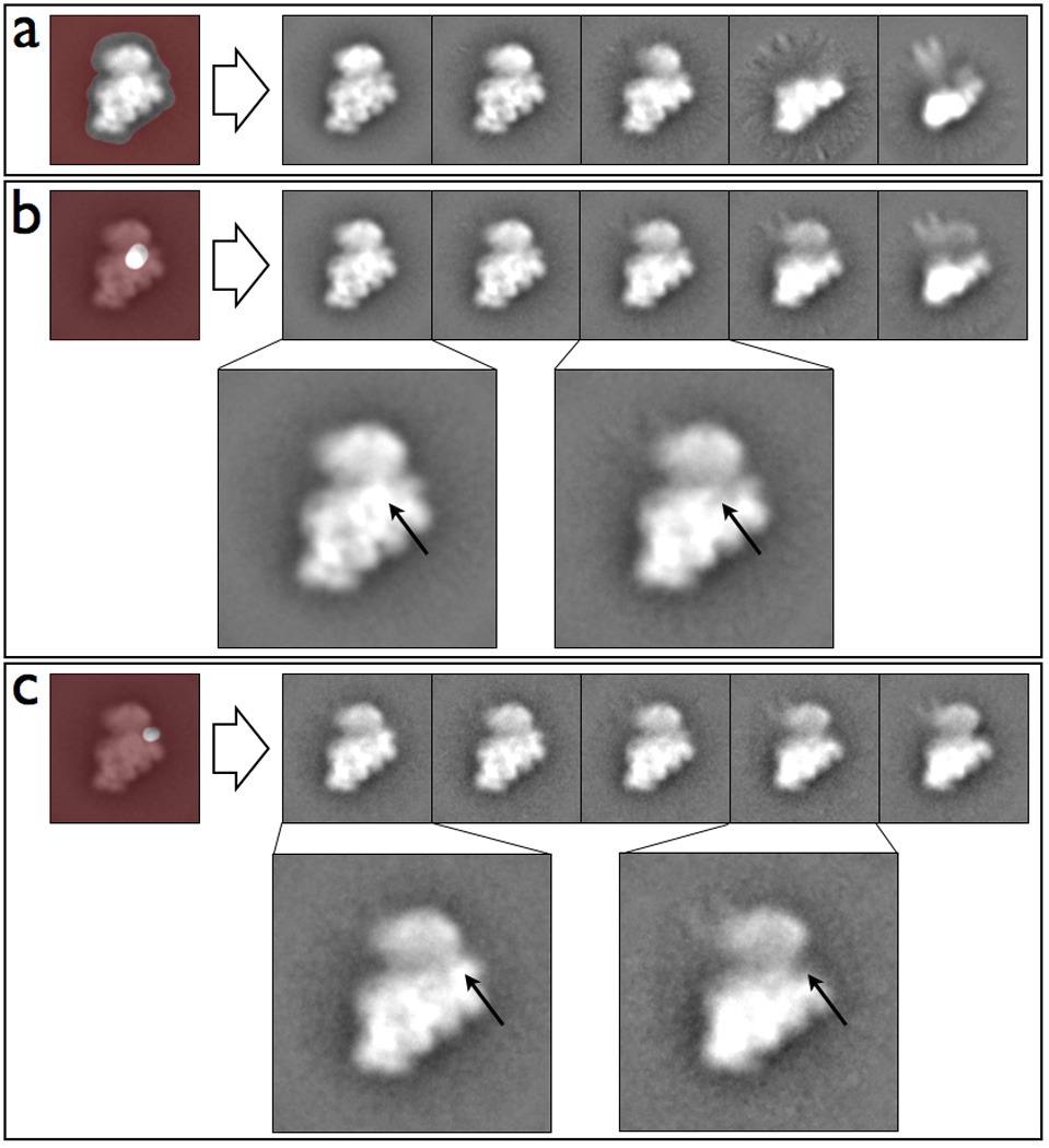
Figure 5.

Classifications of a time point from the in vitro assembly of the 30S ribosomal subunit in Maskiton. (a) Classes resulting from using a mask covering the full subunit, showing that the major variation in the dataset is attributable to unfolding of the 30S ‘head’, and some further unfolding of the 30S ‘body’. Classes resulting from using a mask covering (b) the S2 ribosomal protein and (c) the S11/21 ribosomal protein. (b–c) Arrows in the expanded insets indicate presence / absence of the density corresponding to the masked region.
