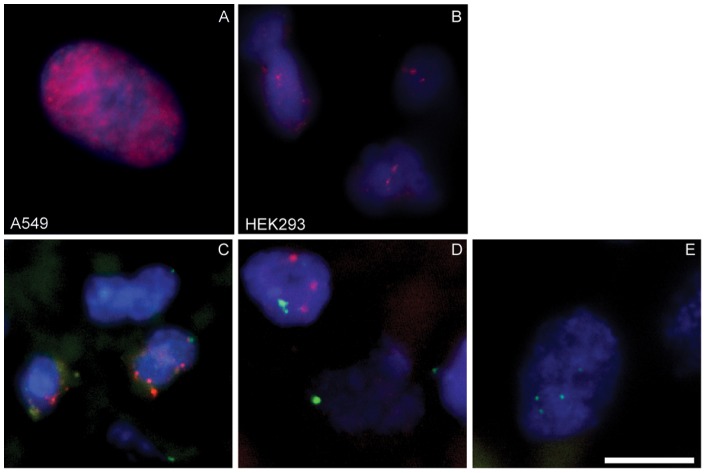
Figure 3. Detection of HAdV by Fluorescence-in-situ-hybridization (FISH).
The hybridization was performed in all experiments with a biotinylated adenovirus DNA probe. Control hybridization was performed on (A) A549 cells infected with HAdV type 5 (24 h, MOI 50) and (B) HEK293. Paraffin-embedded human sarcoma tissue sections were additionally hybridized for the internal positive control (DIG labelled centromeric probe q12). FISH results from (C) a PCR positive leiomyosarcoma (sample 51), (D) a liposarcoma (sample 70) and (E) a PCR negative liposarcoma (sample 47) are shown. For the detection of HAdV-DNA the mouse anti-Biotin-Cy3 antibody (Jackson ImmunoResearch) and for the centromeric probe the sheep-anti-digoxigenin-FITC labelled antibody (Roche) have been used. The bar in picture E represents 10 µm.

