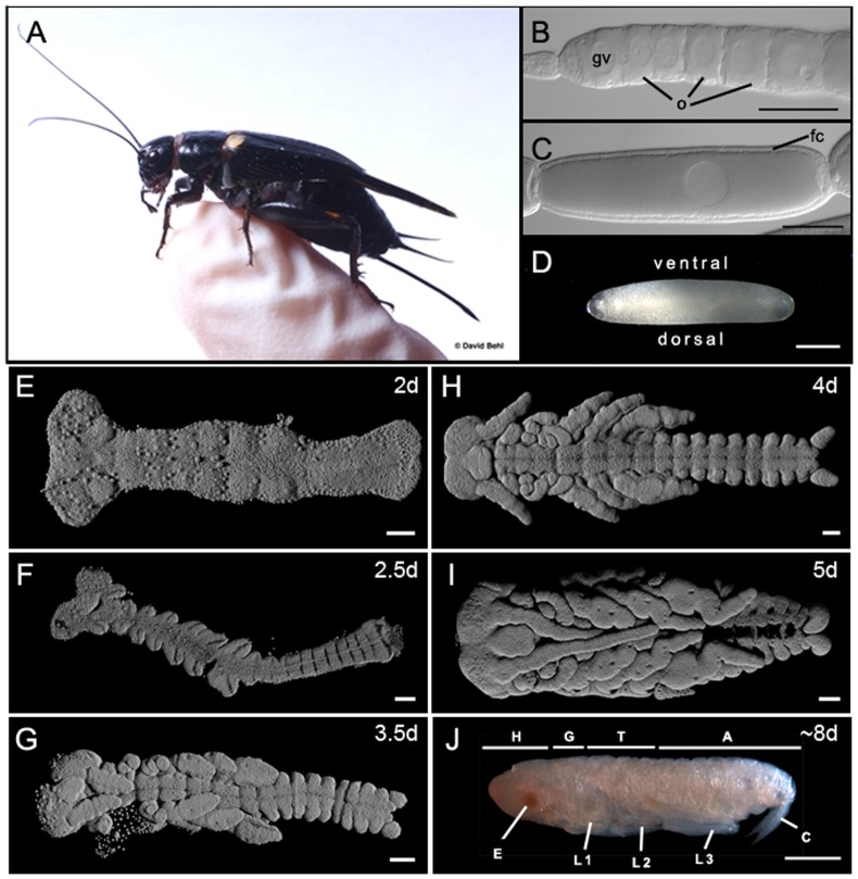
Figure 1. Oogenesis and embryogenesis in the cricket model organism Gryllus bimaculatus.
(A) Adult female cricket perched on a gloved human finger for perspective. (B) Anterior tip of a single ovariole from an adult female ovary, showing oocytes (o) at early previtellogenic stages of oogenesis. A single large germinal vesicle (gv) is distinguishable in each oocyte. Unlike meroistic (containing nurse cells) Drosophila ovaries, G. bimaculatus ovaries are panoistic and lack nurse cells [100]. (C) A single late stage oocyte with a single layer of columnar follicle cells (fc). (D–J) Chronological stages of G. bimaculatus embryogenesis showing the range of embryonic stages represented in the transcriptome presented here. (D) A fertilized egg just after laying. The egg nucleus is distinguishable as a dense patch in the dorsal yolk (arrowhead). Ages are shown as days (d) after egg-laying at 29°C. (E–I) are 3D reconstructions of confocal optical sections of Hoechst 3342-stained embryos dissected free from the egg; (J) is a micrograph of a live embryo dissected free from the chorion. Abbreviations: A = abdomen; C = cerci; E = eye; H = head; G = gnathal segments; L1 = first thoracic leg; L2 = second thoracic leg; L3 = third thoracic leg; T = thorax. Scale bar is 100 µm in (B, C, E–I) and 500 µm in (D, J). Anterior is to the left in all panels. Photo in (A) courtesy of David Behl; photos in (D) and (J) from [101].

