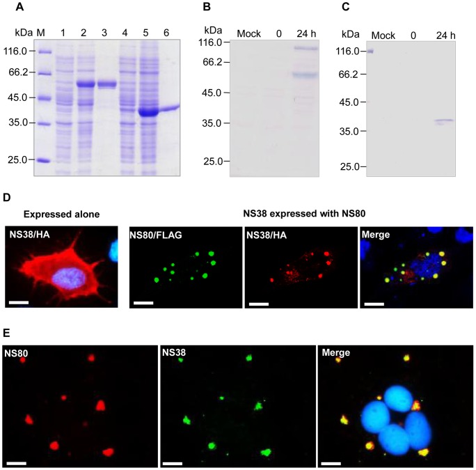
Figure 3. NS80 colocalized with NS38 in transfected and infected cells.
A. Prokaryotic expression of the NS80 and NS38 fusion proteins. M: protein molecular weight marker; lane 1: bacterial lysate containing pET32a-NS80 without IPTG induction; lane 2: bacterial lysate containing pET32a-NS80 with IPTG induction; lane 3: the purified NS80 fusion protein; lane 4: bacterial lysate containing pET28a-NS38 without IPTG induction; lane 5: bacterial lysate containing pET28a-NS38 with IPTG induction; lane 6: the purified NS38 protein. B. Western blot analysis of NS80 expression in infected cells. Two obvious protein bands were detected at 24 h p.i. using anti-NS80 rabbit serum. C. Western blotting analysis of NS38 expression in infected cells. D. In the transfected cells, NS38 (red) is diffusely distributed when expressed alone. However, when NS38 is coexpressed with NS80 (green), they colocalized (yellow). Blue fluorescence indicates the nuclei stained by Hoechst 33342. Bar = 20 μm. E. In infected cells, NS80 (red) and NS38 (green) colocalized in viral factories. Blue fluorescence shows the nuclei stained by Hoechst 33342. Bar = 20 μm.

