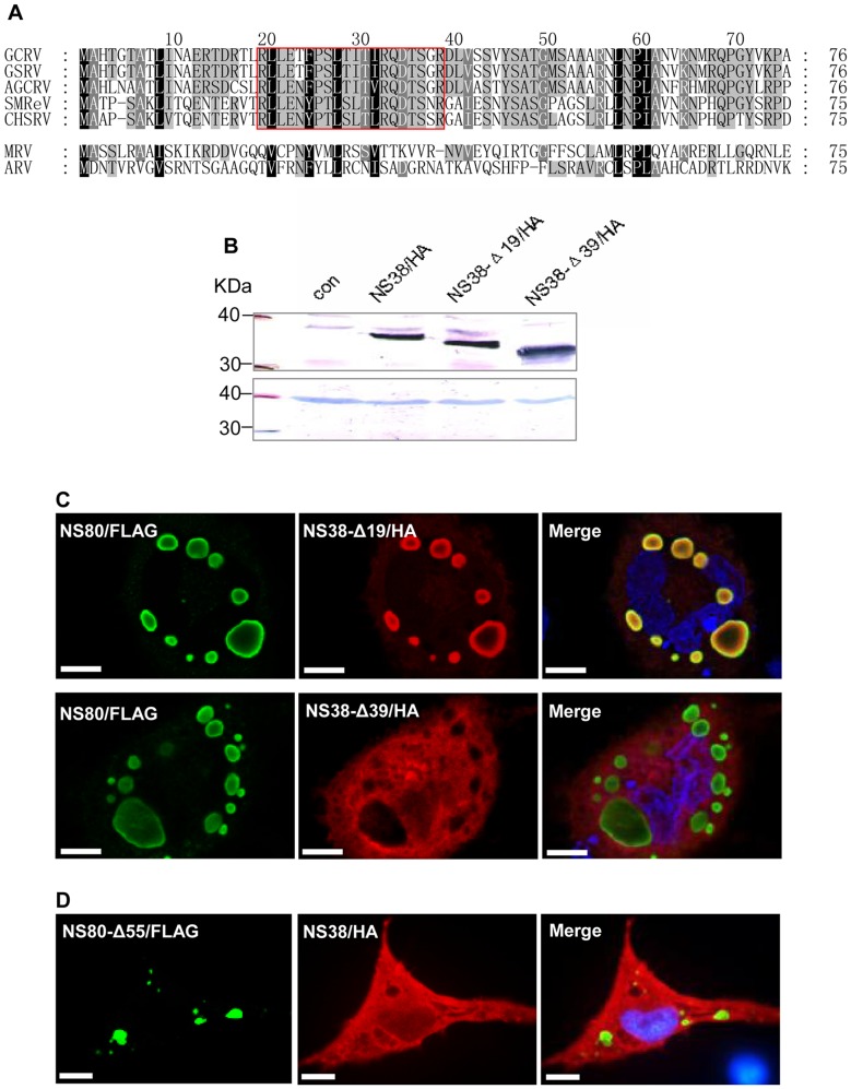
Figure 4. Amino acid sequence comparison of NS38 from aquareoviruses and orthoreoviruses, and immunofluorescence analysis of key sequences in interactions between NS80 and NS38.
A. Amino acids alignment of N-terminal sequences of NS38 from five aquareoviruses (GCRV, CSRV, AGCRV, SMReV and CHSRV) and its homologs belonging to orthoreoviruses (σNS in MRV or ARV). The black shaded regions indicate highly conserved residues in all viruses, while the gray shaded regions are partially conserved residues with more than 80% identity. The key region involved in protein interactions is boxed (aa 20–39 of GCRV). B. Western blot analysis of expression products of full-length and truncated NS38 in transfected cells C. Immunofluorescence analysis of NS80 coexpressed with NS38 N-terminal truncations in transfected cells. NS38-Δ19 (red) colocalized with NS80 (green) but NS38-Δ39 (red) was not colocalized with NS80 (green), which indicated that the NS38 conserved residues, 20–39, were responsible for colocalization with NS80. The nucleus was stained by Hoechst 33342 and presented as blue. Bar = 20 μm. D. Immunofluorescence analysis of NS38 coexpressed with NS80 N-terminal truncations in transfected cells. NS80-Δ55 (green) was not colocalized with NS38 (red). The nucleus was stained by Hoechst 33342 and presented as blue. N-terminal residues, 1–55, of NS80 were important for interactions with NS38. Bar = 20 μm.

