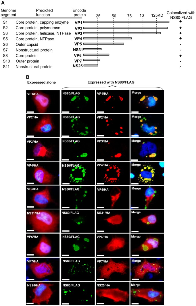
Figure 5. Diagram, summary, and immunofluorescence analysis of interactions between NS80 and the other nine viral proteins.
A. Diagram and summary of interactions between NS80 and the nine viral proteins. “+” indicates that two proteins colocalized, and “−” indicates that two proteins were not colocalized. B. Immunofluorescence analysis of the nine viral proteins when expressed alone or with NS80. The nine viral proteins were expressed alone (lane on the left of the figure) or coexpressed with NS80 (the three lanes on the right of the figure). Green fluorescence indicated NS80. Red fluorescence indicated the other nine viral proteins. The nucleus was stained by Hoechst 33342 and presented as blue. NS80 colocalized with VP1, VP2, VP3, and VP4, and NS80 weakly colocalized with VP6. Bar = 20 μm.

