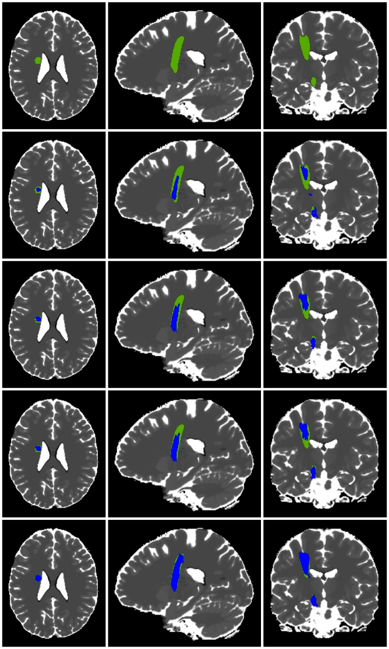
Figure 5. Comparison of two-ROI-approach, whole brain tractography and repeated fiber tracking (example).
Comparison of fiber tracking results in blue achieved by the two-ROI-approach (row 2), traditional whole brain tractography (row 3), variant of whole brain tractography (row 4) and the repeated tracking approach (row 5) in axial, sagittal and coronal view within the anatomical phantom data set and underlying modeled ground truth (row 1) in green.

