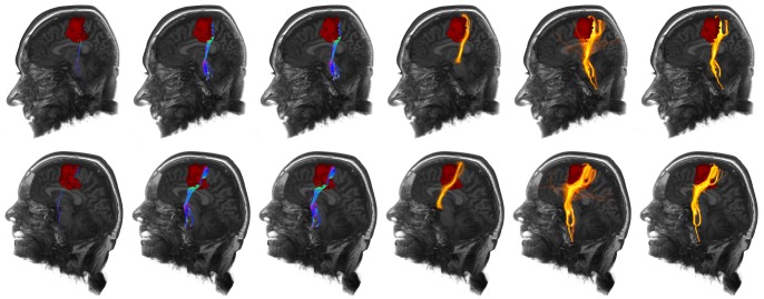
Figure 9. Application of repeated fiber tracking on patient data with a left precentral glioma: corticospinal tract.
Comparison (from left to right) of the results from standard deterministic fiber tracking, variant of whole brain fiber tracking, whole brain fiber tracking, probabilistic fiber tractography, unfiltered results of the repeated fiber tracking method and filtered results of the repeated fiber tracking method with a fiber bundle membership of 50% using a seed region scaling of 2 mm and 128 generated seed regions, tumor segmented manually in red.

