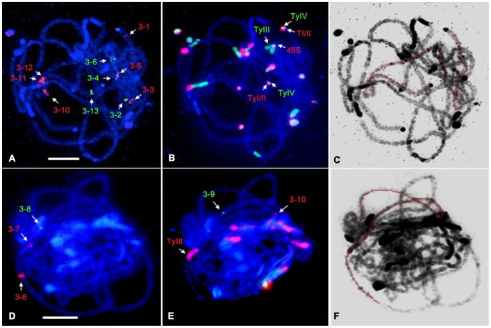
Figure 2. FISH mapping of cucumber chromosome 3-specific fosmid clones and satellite DNAs on the pachytene chromosome 3.
A Fish mapping of 10 fosmid clones on the pachytene chromosome 3 of cucumber. B Fish mapping of 45S rDNA (red), Type I/II (red), Type III (green), and Type IV (green) clones on the same slide as A using reprobing method. C The DAPI-stained chromosomal image was converted as a black–white image to enhance the visualization of chromosome structure. D Fish mapping of 3 fosmid clones on the pachytene chromosome 3 of cucumber. E Fish mapping of Type III (red) and two fosmid clones on the same slide as D using reprobing method. F The DAPI-stained chromosomal image was converted as a black–white image to enhance the visualization of chromosome structure. The pachytene chromosome 3 was orientated with red line. Bars = 5 µm.

